Rendering
To facilitate inspecting segmentation results, Ais comes with a built-in isosurface renderer that is similar to the surface renderer in ChimeraX. As soon as an Export job has created a segmentation, these results are available in the 'Render' tab of the main menu.
By default, Ais looks for segmentation results in the same folder in which the original .mrc file is located. Specify a different directory by clicking the '...' button or editing the path in the 'Volumes' tab of the main menu.
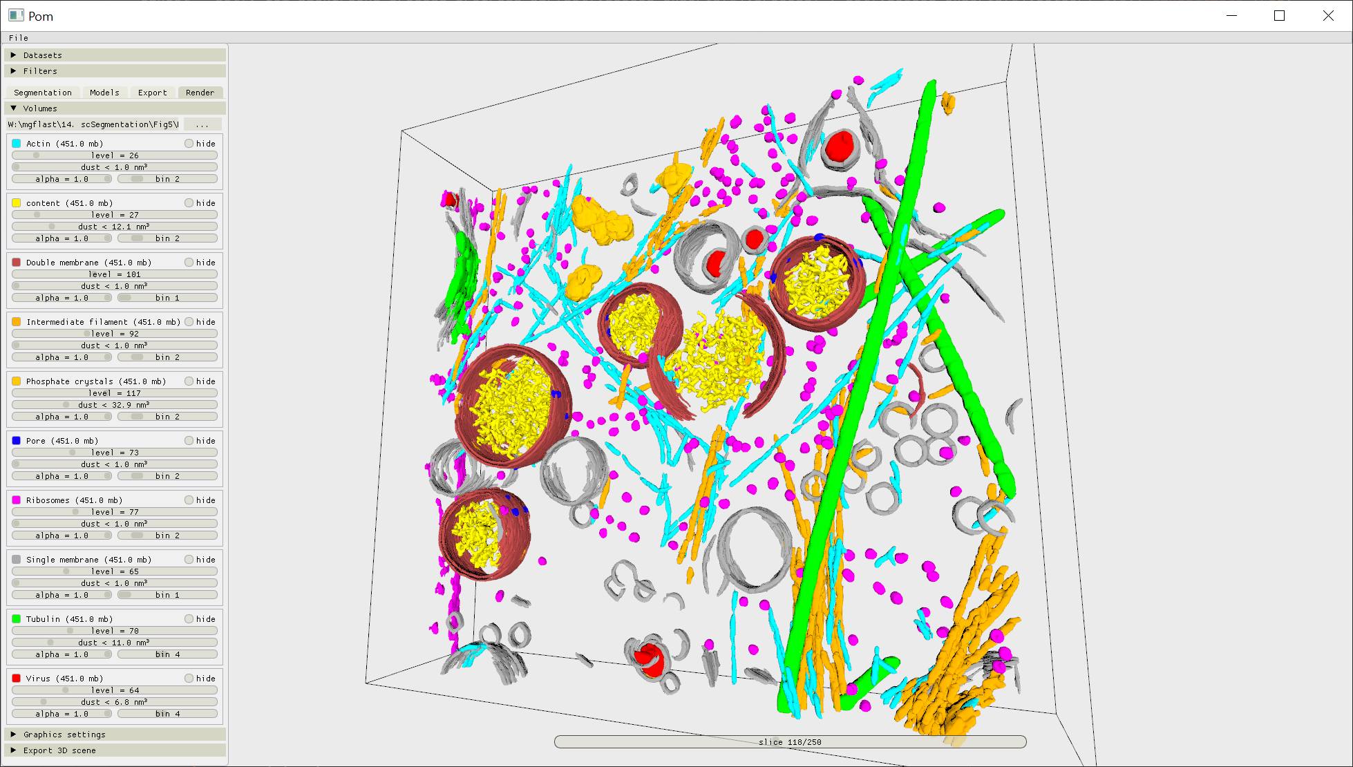
3D models can be ported from Ais directly into ChimeraX, Blender, or saved using the .obj file format, using the buttons in the 'Export 3D scene' submenu.
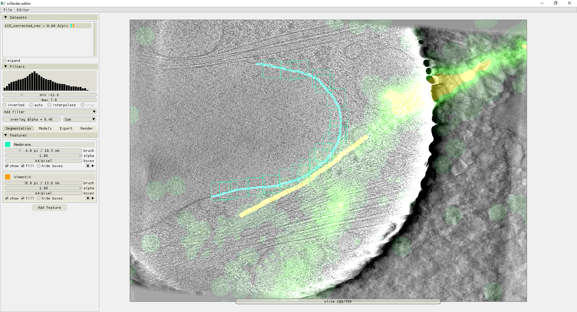
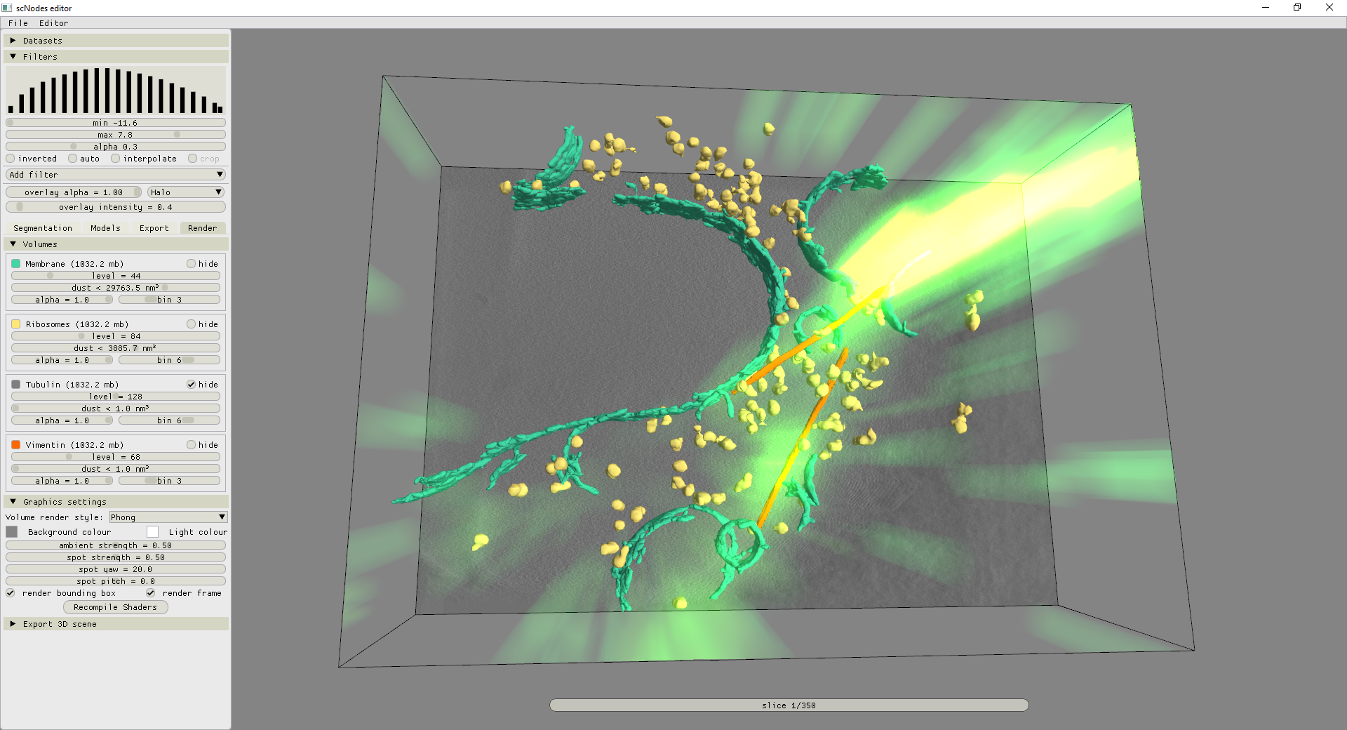
In scNodes+Ais - the version of Ais integrated into our main superCLEM software suite, available at github.com/bionanopatterning/scNodes - additional functionality is available to render fluorescence overlays in the segmentation editor. Tomograms with fluorescence overlays can be prepared in the scNodes' Correlation Editor and can be directly forwarded into Ais with a single button click. These overlays can then be consulted during every step of the segmentation workflow.