3. Actin filaments
Example 3: actin filaments in ghost cells
In this example we used easymode, Pom, Relion5, and M to reconstruct, denoise, segment, pick, and average actin filaments in human immortalized skin fibroblast 'ghost cells' (plasma membrane removed by surfactant treatment).
Dataset and computational resources
For this test we used 143 tilt series of FIB-milled ghost cells which we collected ourselves. They are not yet available online.
We used 4 NVIDIA RTX 4090 GPUs for most processing steps.
At the onset the data consisted of EER frames and mdocs in a single directory. Pixel size was 1.56 Å/px, dose-symmetric tilt series of ±60° with 3° increments and a total dose of 100 electrons/Ų.
project_root/
└── raw/            # .mdoc and .eer files
    ├── Position_2_2_001_0.00_20260324_005054_EER.eer
    ├── Position_2_2_002_3.00_20260324_005145_EER.eer
    ├── Position_2_2.mdoc
    └── ...
Step 1: tomogram reconstruction
easymode reconstruct --frames raw --mdocs raw --no_halfmaps
warp_tiltseries/reconstruction/.
Step 2: tomogram denoising
easymode denoise --data warp_tiltseries/reconstruction --output denoised --method n2n --gpu 0,1,2,3
denoised/.
Step 3: actin segmentation
easymode segment actin --data denoised --output segmented --gpu 0,1,2,3
Step 4: subset selection in Pom
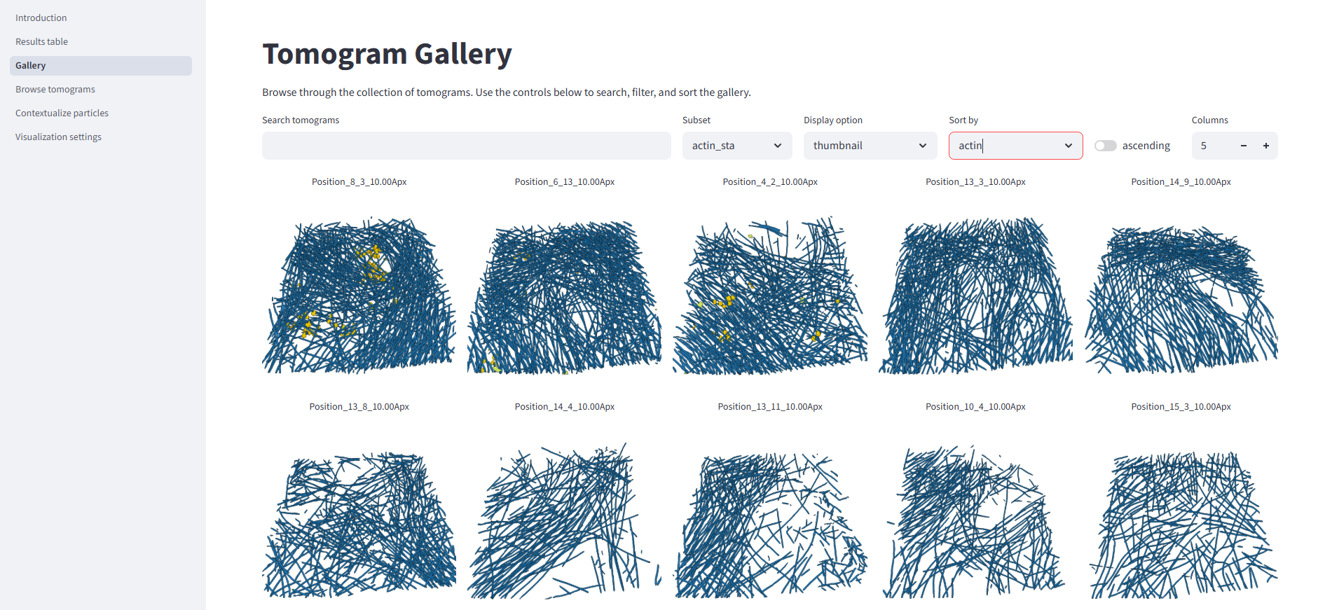
We ran Pom to browse the tomograms and segmentation results. The dataset contained so much actin that we did not need all of it -- we selected a subset of 24 high-contrast tomograms, which we called 'actin_sta' in Pom.
Step 5: filament picking
easymode pick actin --data segmented --output coordinates/actin --filament --spacing 150 --subset pom/subsets/actin_sta.txt
--subsets pom/subsets/actin_sta.txt ensures that we pick particles only in the subset of tomograms that we just created.
In a more difficult case (cellular lamella, rather than ghost cells, for example) it may be worth doing per-filament averaging or an initial round of 3D classification with alignment to discard particles that do not align well or are not really actin. In samples with high thickness or tilt series acquired with a relatively low dose, the actin segmentation outputs tend to under-annotate and under-pick actin.
Step 6: export and initial refinement
WarpTools ts_export_particles --input_directory coordinates/actin --input_pattern "*.star" --coords_angpix 10.0 --output_star relion/actin/particles.star --output_angpix 3.0 --box 80 --diameter 100 --2d --relative_output_paths
Step 7: refinement in M
Refinement in M with refine_particles, refine_imagewarp (up to 4x4), and ctf_defocus + ctf_defocusexhaustive brought the resolution to 3.9 Å.
Subtomogram average at 3.9 Å global resolution, obtained with easymode segment actin, easymode pick actin --filament, and averaging in Relion5/M.